Diametros MedicalHead Office: via Fra’ Domenico Buonvicini 17, 50132 Firenze
Operational Office: via di Terranuova 48/5, 52025 Montevarchi (AR)

Lobster Project® is an implantable device for percutaneous interspinous distraction.
The degenerative stenosis of the lumbar spinal canal is a pathologic disease which leads to the progressive closure of the vertebral foramen and / or the conjugate foramen.
Lobster Project® is an implantable device for percutaneous interspinous distraction.
It is implanted through a percutaneous surgical technique with a specific and dedicated instruments set; it’s indicated for patients with less severe stenosis mainly tied to soft tissues, soft stenosis, or in patients who are not candidates for a surgical interventions under general anesthesia.

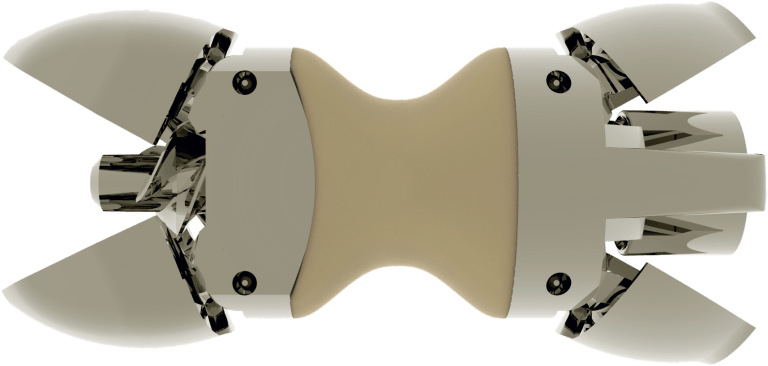
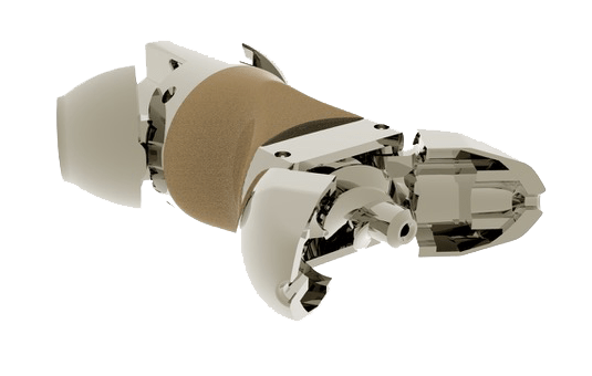
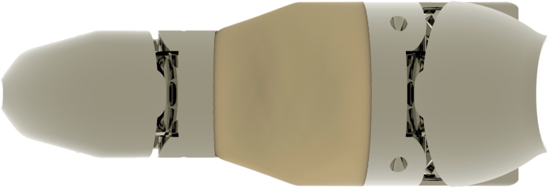
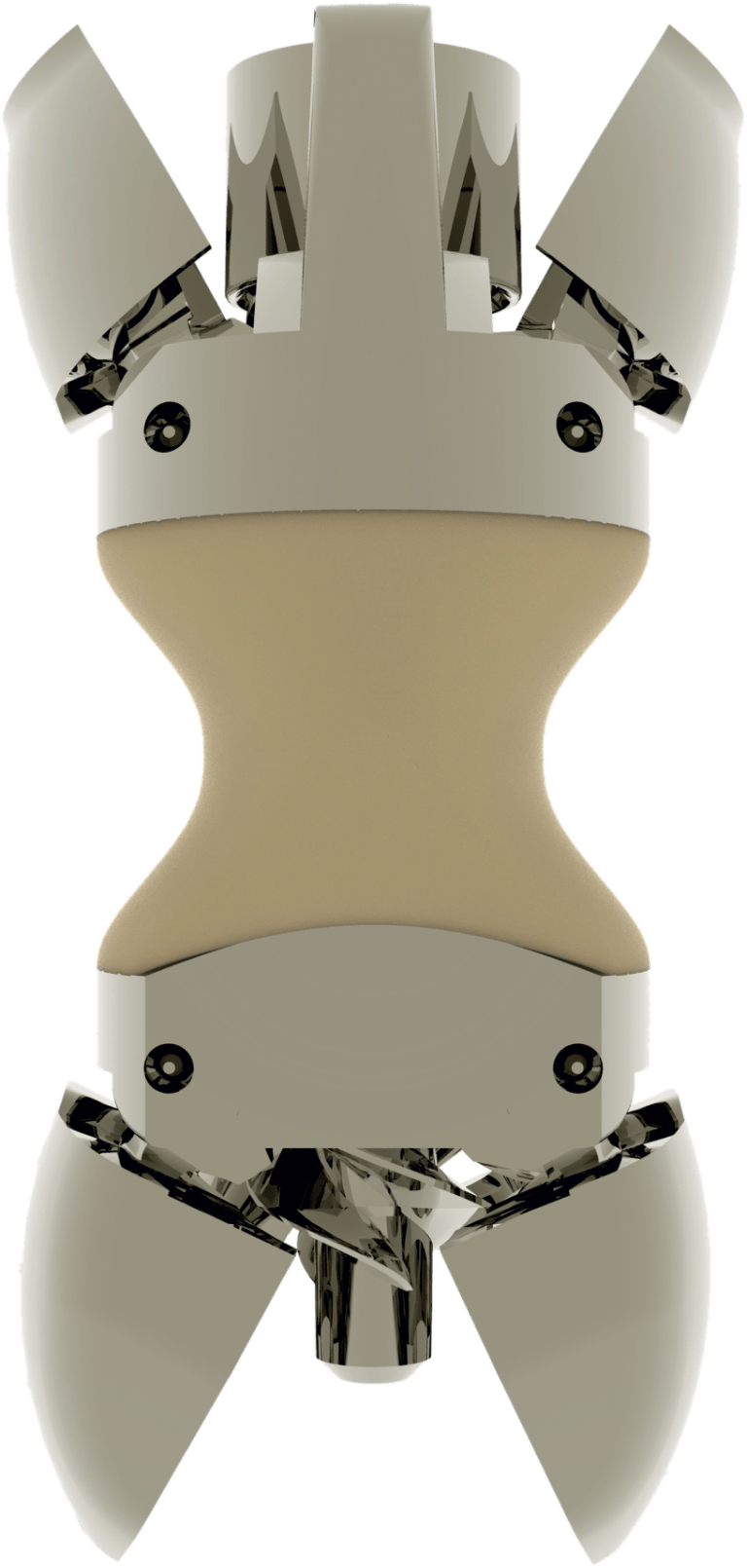
The distance existing between the conservative techniques and the “open” surgery finalized to spinal fusion has been filled with Lobster®, the first dynamic percutaneous interspinous spacer, optionally removable after being implanted. The device is indicated in the treatment of degenerative lumbar canal stenosis, which causes dull ache or acute back pain, which may radiate to the lower limbs with weakness in the legs and loss of balance during walking (claudication neurological).


Through mini-invasive percutaneous technique, the Lobster® device allows the indirect decompression of the spinal canal; it determines the reduction of the freedom of movement in extension of the treated vertebral bodies while keeping the normal rotation capacity and spinal flexion axis. This allows the distension of the posterior portion of the column reducing the eventual disc protrusion and the thickness of the yellow ligaments. These effects increase the size of the spinal canal with reduction or resolution of the associated pain.

How it works

The Lobster® device is intended to stop the segmental extension and to distract the interspinous space between the lumbar spine vertebrae (L1-L5), maintaining the foraminal height, opening up the area of the spinal canal, reducing stress on the facet joints and relieving pressure on the posterior annulus of the intervertebral disc.

Lobster® is implanted through a minimally invasive surgical technique with a specific and dedicated instruments set.
The device consists of a fusiform body with a central saddle, and 4 “wings” that open and close, located at the end of the central body.
The central saddle is designed to be inserted between the two vertebral spinous processes, object of distraction, while long “wings”, opened after the implant placement, prevent ventral and lateral migration of the device.

Benefits:
  • Short surgical procedure under local anesthesia.
  • Percutaneous Technique; small skin incision, less blood loss and reduced muscle trauma. Mini-invasive.
  • Preserves anatomical structures.
Features:
  • Limit Extension movements
  • reduce stress on the facet joints
  • maintain the foraminal height
  • 6 sizes to accommodate different patient anatomies
  • Instrument set allows to close the wings and possibly remove the device
  • 3 different configurations of the device

Configurations:

Lobster Project® is available in 2 different versions:

    1 – Titanium (code LBT)
    2 – Central body in titanium / PEEK coated (code LBS)

All implants are delivered STERILE

Be informed

Certifications:

  • Alexandre A et al. One-year follow-up of a series of 100 patients treated for lumbar spinal canal stenosis by means of HeliFix interspinous process decompression device. Biomed Res Int. 2014;2014:176936.
  • Arnoldi CC, et al. Lumbar spinal stenosis and nerve root entrapment syndromes. Definition and classification. Clin Orthop 1976;115:4–5.
  • Aryanpur J, Ducker T. Multilevel lumbar laminotomies: an alternative to laminectomy in the treatment of lumbar stenosis. Neurosurgery 1990;26:429-32; discussion 33.
  • Bartels RH. The X STOP device. J Neurosurg Spine 2007;6:620-1.
  • Bridwell KH. Lumbar spinal stenosis. Diagnosis, management, and treatment. Clin Geriatr Med. 1994 10(4):677-701.
  • Chiu JC. Interspinous process decompression (IPD) system (X-STOP) for the treatment of lumbar spinal stenosis. Surg Technol Int 2006;15:265-75.
  • Chou R, et al. Nonsurgical interventional therapies for low back pain: a review of the evidence for an American Pain Society clinical practice guideline. Spine (Phila Pa 1976) 2009;34:1078-93.
  • Circ I, et al. The lateral recess syndrome. J Neurosurg 1980;53:433–43.
  • Cooper G.,MD. Foot Drop Symptoms, Steppage Gait & Other Warning Signs. Chp 2. Spine-health 2009.
  • Cuckler J, et al. The use of epidural steroids in the treatment of lumbar radicular pain. J Bone Joint Surg 1985;67A:63–6.
  • Daniel C, et al. A Comparison of Physical Therapy, Chiropractic Manipulation, and Provision of an Educational Booklet for the Treatment of Patients with Low Back Pain. N Engl J Med 1998; 339:1021-1029.
  • Epstein BS,et al. Anatomicoradiological correlations in cervical spine discal disease and stenosis. Clin Neurosurg 1978;25:148-73.
  • Fischgrund JS, et al. Degenerative lumbar spondylolisthesis with spinal stenosis: a prospective, randomized study comparing decompressive laminectomy and arthrodesis with and without spinal instrumentation. Spine (Phila Pa 1976) 1997;22:2807-12.
  • Fox MW, et al. Clinical outcomes and radiological instability following decompressive lumbar laminectomy for degenerative spinal stenosis: a comparison of patients undergoing concomitant arthrodesis versus decompression alone. J Neurosurg 1996;85:793-802.
  • Fox MW, Onofrio BM. Indications for fusion following decompression for lumbar spinal stenosis. Neurosurg Focus 1997;3(2):e2.
  • Guizzardi and Petrini. Interspinous versus interlaminar devices in DDD: Biomechanics tests. international congress center DWG, 2009.
  • Haake M et al. German Acupuncture Trials (GERAC) for Chronic Low Back Pain. Randomized, Multicenter, Blinded, Parallel-Group Trial With 3 Groups. Arch Intern Med. 2007;167(17):1892-1898.
  • Hanraets PR. [Radicular syndrome and low-back pain]. Folia Psychiatr Neurol Neurochir Neerl 1953;56(1):3-20.
  • Herkowitz HN, Kurz LT. Degenerative lumbar spondylolisthesis with spinal stenosis. A prospective study comparing decompression with decompression and intertransverse process arthrodesis. J Bone Joint Surg [Am] 1991;73-A:802-8.
  • Johansson CB et al., Quantitative Comparison of Machined Commercially Pure Ti and Ti-6Al-4V Implant in Rabbit. J. Oral Maxillofac. Implants, 13 (1998), p. 315.
  • Johansson CB. On Tissue Reactions to Metal Implants (PhD thesis, Dept. of Biomaterials/Handicap Research, University of Göteborg, Sweden, 1991).
  • Jonsson B, et al. A prospective and consecutive study of surgically treated lumbar spinal stenosis. Part I: Clinical features related to radiographic findings. Spine (Phila Pa 1976) 1997;22:2932-7.
  • Kabir SM, et al. Lumbar interspinous spacers: a systematic review of clinical and biomechanical evidence. Spine (Phila Pa 1976). 2010 Dec 1;35(25):E1499-506.
  • Kalichman L, et al. Spinal stenosis prevalence and association with symptoms: the Framingham Study. Spine J. 2009;9:545-50
  • Katz JN, et al. Differences between men and women undergoing major orthopedic surgery for degenerative arthritis. Arthritis Rheum 1994;37:687-94.
  • Kenneth P, et al. Lumbar spinal stenosis: anatomy and phatogenesis. Phys. Med Rehabil. Clin. N. Am. 2003;14:1-15.
  • Khoo LT, Fessler RG. Microendoscopic decompressive laminotomy for the treatment of lumbar stenosis. Neurosurgery 2002;51(5 Suppl):S146-54.
  • Kirkaldy-Willis WH, et al. Lumbar spinal stenosis. Clin Orthop 1974;99:30-50.
  • Kluger J. “3-D Bioprinter.” Time. 11 Nov. 2010.
  • Lin WS et al. Additive manufacturing technology (direct metal laser sintering) as a novel approach to fabricate functionally graded titanium implants: preliminary investigation of fabrication parameters.Int J Oral Maxillofac Implants. 2013, 28(6):1490-5.
  • Lingreen R, Grider JS. Retrospective review of patient self-reported improvement and post-procedure findings for mild® (minimally invasive lumbar decompression). Pain Physician. 2010;13(6):555-60.
  • Malmivaara A, et al. Surgical or nonoperative treatment for lumbar spinal stenosis? A randomized controlled trial. Spine (Phila Pa 1976) 2007;32:1-8.
  • Markman JD and Gaud KG. Lumbar spinal stenosis in older adults: current understanding and future directions. Clinics in Geriatric Medicine. 2008; 24:369–388, 2008.
  • Marshall RW. Focus on Understanding and treating spinal stenosis. Journal of Bone and Joint Surgery. 2010.
  • Matsumoto M, et al. Nocturnal leg cramps: a common complaint in patients with lumbar spinal canal stenosis. Spine. 2009;34(5):E189–94.
  • National Institute for Health and Clinical Excellence (NICE). IPG365. Interspinous distraction procedures for lumbar spinal stenosis causing neurogenic claudication. November 2010.
  • Peter F. Ullrich, Jr., MD. Lumbar Spinal Stenosis: A Definitive Guide. Spine – health. 1999 Chp. 1.
  • Podichetty VK, et al. Effectiveness of salmon calcitonin nasal spray in the treatment of lumbar canal stenosis: a double-blind, randomized, placebo-controlled, parallel group trial. Spine (Phila Pa 1976) 2004;29:2343-9.
  • Postacchini F, et al. The surgical treatment of central lumbar stenosis. Multiple laminotomy compared with total laminectomy. J Bone Joint Surg [Br] 1993;75:386-92.
  • Postacchini F.Management of lumbar spinal stenosis. J Bone Joint Surg [Br] 1996;78-B:154-64.
  • Rajagopal TS, et al. Improved diagnosis of spinal stenosis with coronal MRI, 2010 – Research in Progress.
  • Sanderson PL, Wood PL. Surgery for lumbar spinal stenosis in old people. J Bone Joint Surg [Br] 1993;75- B:393-7.
  • Senegas J. Surgery of the intervertebral ligaments, alternative to arthrodesis in the treatment of degenerative instabilities. Acta Orthop Belg 1991;57(suppl 1):221-6.
  • Simmons JW. Posterior lumbar interbody fusion with posterior elements as chip grafts. Clin Orthop Relat Res. 1985;193:85–89.
  • Spratt KF, et al. A predictive model for outcome after conservative decompression surgery for lumbar spinal stenosis. Eur Spine J 2004;13:14-21.
  • Surace MF. Lumbar spinal stenosis treatment with aperius perclid interspinous system. Eur Spine J (2012) 21 (Suppl 1):S69–S74.
  • The spine market group. Spine news, 2013. http://www.thespinemarketgroup.com.
  • Thomas NW, et al. Quantitative outcome and radiographic comparisons between laminectomy and laminotomy in the treatment of acquired lumbar stenosis. Neurosurgery 1997;41:567-74; discussion 74-5.
  • Traini et al. Direct laser metal sintering as a new approach to fabrication of an isoelastic functionally graded material. Dental material 2008, 24:1525-1533.
  • Weinstein JN, et al. Surgical compared with nonoperative treatment for lumbar degenerative spondylolisthesis: four-year results in the Spine Patient Outcomes Research Trial (SPORT) randomized and observational cohorts. J Bone Joint Surg Am 2009;91:1295-304.
  • Weinstein JN, et al. Surgical versus nonsurgical therapy for lumbar spinal stenosis. N Engl J Med 2008;358:794-810.
  • Wu AM et al. Interspinous spacer versus traditional decompressive surgery for lumbar spinal stenosis: a systematic review and meta-analysis. PLoS One. 2014 May 8;9(5):e97142.
  • Zingg PO, Boos N. Lumbar Spinal Stenosis. In: Boos N, Abei, M (eds). Spinal Disorders. Berlin Heidelberg: Springer-Verlag, 2008:513-37. invasive surgical technique with a specific and dedicated instruments set. The device consists of a fusiform body with a central saddle, and 4 “wings” that open and close, located at the end of the central body. The central saddle is designed to be inserted between the two vertebral spinous processes, object of distraction, while long “wings”, opened after the implant placement, prevent ventral and lateral migration of the device.

The information on this site is reserved exclusively for doctors, pharmacists and other health professionals.